基于深度学习并利用时间信息在X射线血管造影中进行冠状动脉血管分割|文献速递-深度学习医疗AI最新文献
Title
题目
Deep learning based coronary vessels segmentation in X-ray angiographyusing temporal information
基于深度学习并利用时间信息在X射线血管造影中进行冠状动脉血管分割
01
文献速递介绍
有创冠状动脉造影(ICA)在冠状动脉疾病(CAD)的诊断和介入治疗的临床实践中被广泛应用(拉什加里等人,2024年)。它是诊断冠状动脉狭窄的金标准,同时为治疗提供定性和定量的指导(科奇卡,2015年)。冠状动脉血管的分割能够实现清晰的可视化,并可计算诸如血流储备分数等定量临床指标(屠等人,2014年)。在有创冠状动脉造影中,获取的血管结构总是与背景结构重叠。图像还会受到身体运动和造影剂分布不均的影响,从而导致分割伪影,如边界错位和血管不连续(布隆代尔等人,2006年)。 在这项工作中,我们应对了这些挑战,并开发出一种完全自动化的方法,以准确有效地从连续的有创冠状动脉造影图像序列中勾勒出冠状动脉血管。 血管分割在文献中受到了大量关注。传统方法包括血管增强和血管跟踪技术(弗兰吉等人,1998年;法兹拉利等人,2015年;耶尔曼等人,2016年),这些方法在存在大规模和对比度变化的情况下常常失效,并且会将有创冠状动脉造影中的其他结构(如导管或肋骨)错误标记。跟踪算法更常用于利用拓扑信息和先验信息对血管中心线进行处理,并且对有创冠状动脉造影的伪影很敏感(邹等人,2009年;安布罗西尼等人,2015年)。 申等人(2016年)和夏等人(2020年)提出了涉及优化和矩阵分解的复杂非机器学习方法。尽管他们的结果可与当前最先进水平(SOTA)相媲美,但诸如测试期间标准差较大以及由于血管不连续导致拓扑结构不完整等问题仍未得到解决。 机器学习方法,尤其是神经网络,已成为包括分割在内的大多数医学图像分析应用中的最先进技术。它们通常涉及U-Net(龙内贝格尔等人,2015年)或全卷积网络(FCN)(朗等人,2015年)的变体。血管分割受到最多关注的领域是视网膜成像(卡姆兰等人,2021年;谭等人,2022年)。马尼尼斯等人(2016年)表明,自动化方法可以实现与人类专家标注相当的视网膜血管勾勒。然而,由于有创冠状动脉造影存在独特的挑战,如灰度成像、不规则的血管结构、血管直径的显著变化以及运动伪影,视网膜血管分割算法不能直接应用于有创冠状动脉造影。此外,对整个血管树,尤其是细小血管进行手动标注既耗时又困难,这是由于人为错误、时间限制以及准确勾勒细小且低对比度血管的固有难度所致。这些因素常常导致标注较为粗略,并使数据集的规模和质量成为提高有创冠状动脉造影分割性能的关键挑战。第二个主要挑战是有创冠状动脉造影图像的质量问题,不一致的对比度常常使血管难以与背景区分开来。诸如造影剂注射时间、血流速度、患者运动以及冠状动脉内造影剂弥散的变化等因素导致了这种不一致性,使准确分割变得更加复杂。最后一个挑战与维持生成的血管树的结构完整性有关,其中尽量减少不连续的血管并避免血管边界的过度或欠分割对于降低狭窄误判的风险至关重要。因此,需要设计特定的解决方案。 1.1 相关工作 纳斯尔 - 伊斯法哈尼等人(2018年)提出了一种冠状动脉血管分割流程,其中包括一个图像增强模块、一个用于上下文特征提取的网络以及一个用于概率特征提取的网络。蒋等人(2021年)开发了一种多尺度、多分辨率的方法,以处理有创冠状动脉造影中冠状动脉血管的对比度分布和尺寸的较大变化。这些带有专门组件以提升性能的方法在主要血管上效果显著,但常常无法区分细小血管,并且无法获得准确的血管边界,这严重影响了狭窄的识别。因此,奥等人(2018年)和杨等人(2019年)开发了仅专注于准确分割单个主要血管(右冠状动脉(RCA)或左前降支(LAD)动脉)或进行狭窄分类的方法。这些方法的一个优点是每张图像仅需要对一个分支进行手动标注,这在相对较大的数据集里是可行的,因为可用的标注有创冠状动脉造影数据集的规模常常限制了冠状动脉血管分割的性能。例如,在纳斯尔 - 伊斯法哈尼等人(2018年)的方法中,金标准仅包含44个有创冠状动脉造影图像。在最近的一项研究中,郝等人(2020年)提出了一种用于冠状动脉血管分割的深度通道注意力网络,使用了一个包含300多个有创冠状动脉造影图像的数据集。一种潜在的策略是使用诸如均值教师模型等方法进行半监督学习,该方法可以从未标注的图像中提取有意义的信息(何等人,2022年)。 运动伪影,尤其是来自心脏和呼吸运动的伪影,会对有创冠状动脉造影的质量产生不利影响。伴随着造影剂的冲洗效应,即使是人类专家也难以识别血管。对运动物体分割的早期尝试可以追溯到计算机视觉领域。弗拉基阿达基等人(2015年)提出了一种双通路神经网络,该网络采用RGB视频及其光流来检测视频序列中的物体。弗隆佐斯和米科拉伊奇克(2018年)采用了类似的方法,在低级二进制分割和光流上使用了一个多阶段U-Net架构。郝等人(2020年)采用了带有时间融合卷积和通道注意力的传统U-Net,但并未旨在维持血管树的结构完整性。梁等人(2021年)设计了半三维U-Net,该网络结合了时间特征提取,用于从血管造影视频中分割冠状动脉血管,重点是通过利用空间和时间特征来提高分割质量。他们的方法只能考虑以目标帧为中心的奇数个时间帧。万等人(2021年)将全卷积网络应用于三帧有创冠状动脉造影分割,使用三通路全卷积网络和影响矩阵来解码时间信息。这些方法常常面临局限性,例如在更密集的网络中缺乏对有效推断局部和全局信息的分析。 类似的方法已被用于数字减影血管造影(DSA)中的脑血管分割。苏等人(2024年)采用了一种时空U-Net,通过时间学习模块同时集成空间和时间特征,从而能够对时空动态进行连贯的解码和学习。相比之下,谢等人(2024年)提出了DSANet,该网络将空间和时间特征提取过程解耦。该模型利用时间变换器模块来捕获时间关系,并使用时空融合模块来合并空间和时间信息,从而产生了一种级联解码方法。然而,与有创冠状动脉造影图像相比,脑血管的数字减影血管造影在背景结构的复杂性和运动伪影方面都有所降低。 解决血管不连续的挑战有助于直接应对有创冠状动脉造影图像质量的挑战,从而提高分割效果。已经有一些针对视网膜图像分割提出的尝试解决血管不连续问题的方法。林等人(2022年)创建了一个类似于生成对抗网络(GAN)的模型,带有一个判别网络来指导分割网络。李等人(2020年)提出了IterNet,该网络通过权重共享和模型内跳跃连接多次迭代一个小型U-Net。这些都是隐式方法,对于改善血管连通性的机制没有明确的解释。兰等人(2020年)设计了一个损失函数,通过将不连续的部分融合在一起来缩小预测的血管边界与金标准边界之间的差距。希特等人(2021年)提出将ClDice损失与Dice损失一起使用,重点是保留分割的管状结构的骨架并确保拓扑准确性,特别是保持连接组件和分支的连续性。然而,它对精确的边界勾勒不太敏感,这可能会影响在诸如狭窄区域等具有临床意义区域的性能。奥纳等人(2022年)和克拉夫等人(2022年)引入了连接优化损失函数,旨在保留特定的结构特征,包括线性连续性、背景区域的分离和全局连接性。虽然这些方法在保持类似网络结构的这些方面有效,但它们优先考虑连接性而非边界精度,这可能限制了它们在具有精细细节和复杂分支模式的冠状动脉血管中的适用性。 最近的研究工作主要集中在自适应或先进的架构上。伊森塞等人(2020年)提出的nnUNet框架已成为医学图像分割任务中广泛采用的基线,这是因为它具有在不同数据集上进行泛化的自适应功能。赵等人(2023年)使用图注意力网络进行基于图的节点相似性比较,用于骨架分割。这种方法侧重于利用节点关系来提高分割准确性。何等人(2024年)融合了图注意力网络和卷积神经网络,以在冠状动脉血管分割过程中学习全局几何信息。此外,阮和向(2024年)引入了视觉曼巴U-Net(VM-UNet),该网络利用状态空间模型(SSMs)以线性计算复杂度捕获广泛的上下文信息和长距离交互。这些方法在各种情况下都非常有效,尽管并非专门为应对有创冠状动脉造影序列的独特挑战而设计。 1.2 贡献 对文献的分析以及我们对郝等人(2020年)中使用的公开的323个有创冠状动脉造影短序列样本的研究结果表明,第二个挑战中的运动伪影是有创冠状动脉造影中冠状动脉血管分割准确性有限的主要原因。身体运动可能会将早期帧中清晰勾勒的血管或部分血管移动到目标帧中的低对比度区域,从而导致血管边界不准确和潜在的不连续性。这种结构完整性的破坏最终加剧了与血管不连续相关的问题。人类标注员通常通过可视化有创冠状动脉造影序列,利用连续帧之间的关系来解决重叠血管的问题,从而减轻这一问题。这表明机器学习方法也可以从观察多个有创冠状动脉造影帧中受益,模仿人类标注员的行为。因此,我们假设引入时间信息是在无需复杂运动建模的情况下解决与运动相关挑战的基础。 我们引入了一种新颖的架构,即时间血管分割网络(TVS-Net),在该模型中,时间被视为第三维,能够有效地提取时空特征以应对运动伪影。我们的主要贡献可以总结为四个方面: - 我们开发了一个新的三维(二维+时间)框架,该框架可以同时从多个连续的有创冠状动脉造影帧中提取特征,以分割目标帧。我们的框架结合了一种新颖的密集嵌套三维编码器,该编码器通过其中间层中的额外卷积节点进行扩展,以及一个高度连接的二维解码器。这种双通路设计以UNet++为骨干,确保了强大的时空特征提取和精确的空间识别,从而实现了高保真的分割。 - 我们纳入了一个保持连接性的损失函数(兰等人,2020年),以维持血管结构的完整性,并采用特定的骨架化指标来评估结构准确性。这种方法对于生成用于三维重建的精确血管骨架至关重要。 - 我们的TVS-Net在各种不同的数据源和标注协议下均优于可比方法,在一个公开数据集上实现了83.4%的Dice系数和84.3%的召回率。在一个经过精细标注的精炼子集上,它实现了更高的86.3%的召回率,显著超过了原始数据集的手动标注结果。 - TVS-Net在一个由60个有创冠状动脉造影图像组成的外部数据集上达到了78.5%的Dice系数和82.4%的召回率,超过了所有当前最先进的方法。这些结果突出了我们方法的鲁棒性和泛化性。
Aastract
摘要
Invasive coronary angiography (ICA) is the gold standard imaging modality during cardiac interventions.Accurate segmentation of coronary vessels in ICA is required for aiding diagnosis and creating treatment plans.Current automated algorithms for vessel segmentation face task-specific challenges, including motion artifactsand unevenly distributed contrast, as well as the general challenge inherent to X-ray imaging, which is thepresence of shadows from overlapping organs in the background. To address these issues, we present TemporalVessel Segmentation Network (TVS-Net) model that fuses sequential ICA information into a novel denselyconnected 3D encoder-2D decoder structure with a loss function based on elastic interaction. We develop ourmodel using an ICA dataset comprising 323 samples, split into 173 for training, 82 for validation, and 68for testing, with a relatively relaxed annotation protocol that produced coarse-grained samples, and achieve83.4% Dice and 84.3% recall on the test dataset. We additionally perform an external evaluation over 60images from a local hospital, achieving 78.5% Dice and 82.4% recall and outperforming the state-of-the-artapproaches. We also conduct a detailed manual re-segmentation for evaluation only on a subset of the firstdataset under strict annotation protocol, achieving a Dice score of 86.2% and recall of 86.3% and surpassingeven the coarse-grained gold standard used in training. The results indicate our TVS-Net is effective for multiframe ICA segmentation, highlights the network’s generalizability and robustness across diverse settings, andshowcases the feasibility of weak supervision in ICA segmentation
有创冠状动脉造影(ICA)是心脏介入治疗过程中的金标准成像方式。在有创冠状动脉造影中,准确分割冠状动脉血管对于辅助诊断和制定治疗方案是必要的。目前,用于血管分割的自动算法面临着特定任务的挑战,包括运动伪影和造影剂分布不均匀,以及X射线成像所固有的一般性挑战,即背景中存在来自重叠器官的阴影。 为了解决这些问题,我们提出了时间血管分割网络(TVS-Net)模型,该模型将连续的有创冠状动脉造影信息融合到一种新颖的密集连接的三维编码器-二维解码器结构中,并采用基于弹性相互作用的损失函数。我们使用一个包含323个样本的有创冠状动脉造影数据集来开发我们的模型,将其分为173个用于训练、82个用于验证和68个用于测试,采用了相对宽松的注释协议,生成了粗粒度的样本,并在测试数据集上达到了83.4%的Dice系数和84.3%的召回率。 此外,我们对来自当地一家医院的60张图像进行了外部评估,实现了78.5%的Dice系数和82.4%的召回率,优于当前最先进的方法。我们还在严格的注释协议下仅对第一个数据集中的一个子集进行了详细的手动重新分割以用于评估,获得了86.2%的Dice系数和86.3%的召回率,甚至超过了训练中使用的粗粒度金标准。 结果表明,我们的TVS-Net对于多帧有创冠状动脉造影分割是有效的,突出了该网络在不同场景下的泛化性和鲁棒性,并展示了在有创冠状动脉造影分割中弱监督的可行性。
Method
方法
This section introduces the input data, describes in detail the neuralnetwork architecture and loss function, and explains post-processing,evaluation methods, evaluation metrics, as well as the experimentalsettings and comparison methods. We denote 𝑋 as the ICA sequence,with 𝑇 being the total number of frames. 𝑥𝑡 is frame number 𝑡, 𝑥𝑡=0is the selected ICA frame for manual labeling, and 𝑦 is gold standardsegmentation, where 𝑋 ∈ [0, 255]𝑇×𝐻×𝑊 and 𝑦 ∈ {0, 1}𝐻×𝑊 with 𝐶, 𝑇 ,𝐻, and 𝑊 representing the number of channels, number of temporalframes, and height and width of the frame, respectively. A full datasetis 𝐷 = {(𝑋𝑛 , 𝑦𝑛 )}𝑁 𝑛=1 with 𝑁 being the total number of cases. Thesegmentation output of the network is 𝑓𝑠 (⋅).
本节介绍了输入数据,详细描述了神经网络架构和损失函数,并解释了后处理、评估方法、评估指标,以及实验设置和对比方法。我们将(X)表示为有创冠状动脉造影(ICA)序列,其中(T)为总帧数。(x_t)是第(t)帧,(x{t = 0})是选定用于手动标注的ICA帧,(y)是金标准分割结果,其中(X\in[0,255]^{T\times H\times W}),(y\in{0,1}^{H\times W}),(C)、(T)、(H)和(W)分别表示通道数、时间帧数以及帧的高度和宽度。完整的数据集为(D = {(X_n, y_n)}{n = 1}^{N}),其中(N)是病例总数。网络的分割输出为(f_s(\cdot))。
Conclusion
结论
In this paper, we develop a novel deep learning framework witha densely connected 3D encoder-2D decoder, named TVS-Net, that utilizes multiple frames of ICA sequences for generating accurate coronaryvessels segmentation. The architecture integrates temporal convolution blocks for fusing the image sequence information and a uniqueenergy loss for enhancing topology preservation onto a dense framework featuring deep supervision. This framework can be combinedwith vascular post-processing techniques, such as the method proposedby Qiu et al. (2023), to further enhance segmentation quality. To enabledirect comparison between our method and the closest SOTA temporalsegmentation (Hao et al., 2020) and other SOTA methods, we trainour framework on the same public dataset using the same data split.The experimental analysis not only demonstrates the advantages of amulti-frame approach but also illustrates the problem of incomplete annotations of vascular trees. The significant performance improvementsobserved by the TVS-Net over UNet++ underscore the effectivenessof integrating densely connected 3D–2D temporal blocks. We proposethe use of skeletonization metrics and demonstrate the ability of ourmethod to preserve thin vascular structures accurately. The combineduse of spatio-temporal encoding, connectivity-preserving loss function,and deep supervision enables our proposed TVS-Net to capture a widerange of vessel appearances and motion patterns. This generalizabilityis further highlighted in our evaluation using data collected from a localhospital, where our method achieves the highest Dice, recall, precision,and AUPRC of 78.49%, 82.41%, 75.79% and 0.8437, respectively,despite the fact that our hyperparameters were optimized for the publicdataset alone and no retraining was performed. The smaller size of theprivate dataset and the random selection of cases may contribute togreater variance and performance variability. Due to the OOD nature ofthe private data, all models show less confident predictions. However,the fact that our method excels in this context demonstrates its abilityto adapt to different annotation protocols and data characteristics andhighlights its strong potential for real-world deployment. Additionally,we proposed a variant of TVS-Net, named TVS-Net+, with the sameideology after expanding the 3D encoder. However, due to the largersize of TVS-Net and available GPU resources, its batch size is limited to4, which impacts the network’s ability to generalize effectively, therebylimiting the observed performance gains of TVS-Net+.We re-segment 10 cases with a strict annotation protocol, includingall visible vessels, and use it as the new (fine-grained) gold standardfor the same evaluation pipeline. Trained by the original (coarsegrained) gold standard, our proposed TVS-Net achieves a recall of86.26%, which is 0.96% higher than the original gold standard and16% higher than the current SOTA method SVS-Net (Hao et al., 2020).The performance in accurately preserving vascular skeletons achieves84.00% in 𝐶**𝑟 , improving on the original gold standard by 9.36% andSVS-Net by 26.54%. The qualitative evaluation illustrates the improvements, including the reduction in over-segmented vascular boundaries.Most interestingly, our extensive analyses demonstrate the feasibility ofweak supervision with coarse-grained annotations for coronary vesselssegmentation. This is evidenced by the superior delineation achievedby the TVS-Net model compared to its training gold standard — thecoarse-grained annotations when evaluated against fine-grained annotations. It is important to note that the ICA datasets were created byselecting high-quality frames, limiting our ability to fully demonstratethe network’s performance across low-quality frames, where manualsegmentation is more challenging and often results in coarser gold standards. Consequently, by modulating the completeness level of manualannotations in the dataset, this framework can also facilitate the exploration of a time-performance trade-off between manual and automaticsegmentations in annotation protocols, as well as elucidate the impactof partially segmented ground truth on final trained segmentationquality
在本文中,我们开发了一种新颖的深度学习框架,即具有密集连接的三维编码器-二维解码器的时间血管分割网络(TVS-Net),它利用有创冠状动脉造影(ICA)序列的多个帧来实现准确的冠状动脉血管分割。该架构集成了时间卷积模块以融合图像序列信息,以及一种独特的能量损失函数,用于在具有深度监督的密集框架上增强拓扑结构的保留。这个框架可以与血管后处理技术相结合,比如邱等人(2023年)提出的方法,以进一步提高分割质量。 为了使我们的方法能够与最相近的当前最先进的时间分割方法(郝等人,2020年)以及其他当前最先进的方法进行直接比较,我们在相同的公开数据集上使用相同的数据划分来训练我们的框架。实验分析不仅展示了多帧方法的优势,还说明了血管树标注不完整的问题。TVS-Net相对于UNet++在性能上的显著提升强调了集成密集连接的三维-二维时间模块的有效性。我们提出使用骨架化指标,并证明了我们的方法能够准确保留细小血管结构的能力。时空编码、保持连接性的损失函数以及深度监督的综合运用,使我们提出的TVS-Net能够捕捉到广泛的血管外观和运动模式。 在我们使用从当地一家医院收集的数据进行的评估中,这种泛化能力得到了进一步凸显。尽管我们的超参数仅针对公开数据集进行了优化且未进行重新训练,但我们的方法在该评估中分别实现了最高的Dice系数(78.49%)、召回率(82.41%)、精确率(75.79%)和曲线下面积(AUPRC,0.8437)。私有数据集规模较小以及病例的随机选择可能导致更大的方差和性能差异。由于私有数据具有分布外(OOD)的性质,所有模型的预测结果都显得信心不足。然而,我们的方法在这种情况下表现出色,这证明了它适应不同标注协议和数据特征的能力,并凸显了其在实际应用中强大的潜力。 此外,我们提出了TVS-Net的一个变体,称为TVS-Net+,它在扩展了三维编码器后采用了相同的理念。然而,由于TVS-Net+的规模较大以及可用的GPU资源限制,其批量大小被限制为4,这影响了网络的有效泛化能力,从而限制了TVS-Net+所观察到的性能提升。 我们按照严格的标注协议对10个病例进行了重新分割,包括所有可见的血管,并将其作为新的(细粒度的)金标准用于相同的评估流程。在原始(粗粒度的)金标准的训练下,我们提出的TVS-Net实现了86.26%的召回率,比原始金标准高0.96%,比当前最先进的方法SVS-Net(郝等人,2020年)高16%。在准确保留血管骨架方面的性能在(C_r)指标上达到了84.00%,比原始金标准提高了9.36%,比SVS-Net提高了26.54%。定性评估展示了这些改进,包括过度分割的血管边界的减少。 最有趣的是,我们广泛的分析证明了对于冠状动脉血管分割而言,使用粗粒度标注进行弱监督的可行性。TVS-Net模型与训练时的金标准(粗粒度标注)相比,在与细粒度标注进行评估时实现了更优的勾勒效果,这就证明了这一点。需要注意的是,有创冠状动脉造影数据集是通过选择高质量的帧创建的,这限制了我们全面展示网络在低质量帧上性能的能力,在低质量帧上手动分割更具挑战性,并且常常会产生更粗糙的金标准。因此,通过调节数据集中手动标注的完整程度,这个框架还可以促进在标注协议中探索手动分割和自动分割之间的时间-性能权衡,以及阐明部分分割的真实标注对最终训练的分割质量的影响。
Results
结果
In this section, we present extensive quantitative and qualitativecomparisons. First, we compare the performance of TVS-Net againstits variant, TVS-Net+, to evaluate architectural effectiveness. Next, webenchmark TVS-Net against SOTA methods, highlighting its superiorperformance. Following this, we assess its generalizability on an outof-distribution (OOD) dataset. Additionally, we evaluate the impact ofour loss function and deep supervision setting on segmentation performance. Finally, we conduct a comprehensive study on the re-segmentedgold standard.
在本节中,我们进行了大量的定量和定性比较。首先,我们将TVS-Net与其变体TVS-Net+的性能进行比较,以评估架构的有效性。接下来,我们将TVS-Net与最先进的(SOTA)方法进行基准测试,突出其优越的性能。在此之后,我们在一个分布外(OOD)数据集上评估其泛化能力。此外,我们还评估了我们的损失函数和深度监督设置对分割性能的影响。最后,我们对重新分割的金标准进行了一项全面的研究。
Figure
图

Fig. 1. The proposed TVS-Net model (left) and TVS-Net+ (right) for vessels segmentation from ICA sequences using temporal information. 3D and 2D blocks are represented inpurple and orange, respectively, with all kernel sizes and strides shown in the boxes. The gray output paths at the top indicate deep supervision.
图1:所提出的用于利用时间信息从有创冠状动脉造影(ICA)序列中进行血管分割的时间血管分割网络(TVS-Net)模型(左)和增强版时间血管分割网络(TVS-Net+)(右)。三维(3D)模块和二维(2D)模块分别用紫色和橙色表示,所有的卷积核大小和步长都标注在方框内。顶部的灰色输出路径表示深度监督。
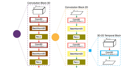
Fig. 2. Details of the convolution blocks and temporal block with the same legend forcolored path in Fig. 1
图2:卷积模块和时间模块的详细信息,其彩色路径的图例与图1中的一致。
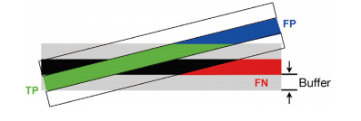
Fig. 3. Skeletonization metrics for vessel centerline with 1-pixel width. True Positives(TP) are green, whereas False Positives (FP) and False Negatives (FN) are blue and red,respectively
图3:宽度为1像素的血管中心线的骨架化指标。真正例(TP)为绿色,而假正例(FP)和假负例(FN)分别为蓝色和红色。
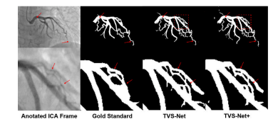
Fig. 4. Qualitative evaluation of segmentation performance using TVS-Net and TVSNet+. The bottom row is a zoomed-in version of the red square in the top row
图4:使用时间血管分割网络(TVS-Net)和增强版时间血管分割网络(TVS-Net+)对分割性能进行的定性评估。底行是顶行中红色方框区域的放大图。
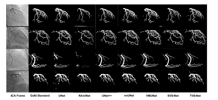
Fig. 5. Qualitative evaluation of segmentation performance of SOTA methods and TVS-Net on dataset 𝐷1 .
图5:在数据集(D_1)上,对当前最先进(SOTA)方法和时间血管分割网络(TVS-Net)的分割性能进行的定性评估。
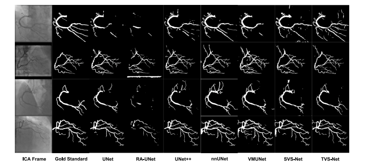
Fig. 6. Qualitative evaluation of segmentation performance of SOTA methods and TVS-Net on OOD dataset 𝐷2
图6:在分布外(OOD)数据集(D_2)上,对当前最先进(SOTA)方法和时间血管分割网络(TVS-Net)的分割性能进行的定性评估。
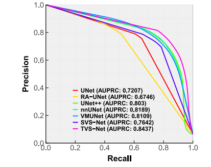
Fig. 7. Precision–Recall curve for evaluation on OOD dataset 𝐷2 .
图7:在分布外(OOD)数据集(D_2)上进行评估的精确率-召回率曲线。
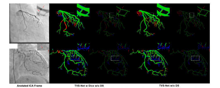
Fig. 8. Qualitative evaluation of segmentation and skeletonization performance of TVS-Net with Dice loss and energy loss, without deep supervision. The color codes for TP, FP,and FN are the same as in Fig. 3.
图8:在无深度监督的情况下,对使用Dice损失函数和能量损失函数的时间血管分割网络(TVS-Net)的分割和骨架化性能进行的定性评估。真正例(TP)、假正例(FP)和假负例(FN)的颜色编码与图3中的相同。
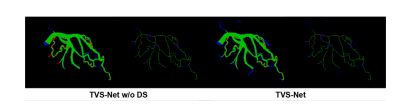
Fig. 9. Vessel segmentation and skeletonization performance of TVS-Net without andwith deep supervision. Color codes are the same
图9:有无深度监督情况下,时间血管分割网络(TVS-Net)的血管分割和骨架化性能。颜色编码相同。
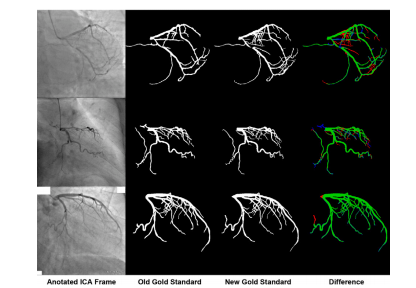
Fig. 10. Three re-segmented samples with minimum (81.61%), median (84.69%), andmaximum (94.76%) Dice scores (top to bottom). Color codes are the same.
图10:三个重新分割的样本,其Dice系数分别为最小值(81.61%)、中位数(84.69%)和最大值(94.76%)(从上至下排列)。颜色编码保持一致。
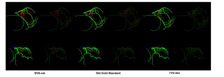
Fig. 11. Qualitative evaluation of segmentation and skeletonization on the new gold standard. The same color code is used here
图11:基于新金标准对分割和骨架化处理的定性评估。此处使用了相同的颜色编码。
Table
表
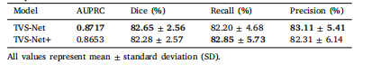
Table 1Comparison of different architecture variants.
表1:不同架构变体的比较。
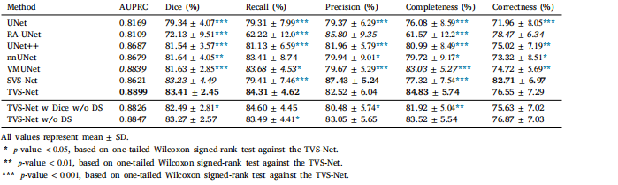
Table 2Comparison of TVS-Net with SOTA methods on test dataset of 𝐷1 .
表2:在数据集(D_1)的测试集上,时间血管分割网络(TVS-Net)与当前最先进(SOTA)方法的比较。
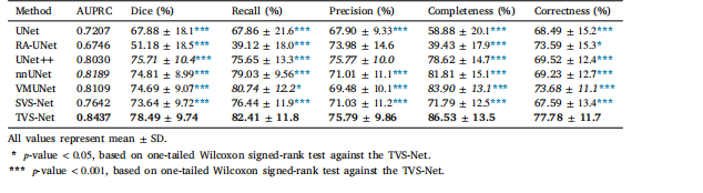
Table 3Comparison of TVS-Net with SOTA methods on OOD dataset 𝐷2 .
表3:在分布外(OOD)数据集(D_2)上,时间血管分割网络(TVS-Net)与当前最先进(SOTA)方法的比较。
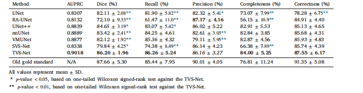
Table 4Performance evaluation on the new gold standard with 10 re-segmented samples.
表4:基于重新分割的10个样本所构成的新金标准进行的性能评估。